On the 14th, Union Hospital affiliated with Tongji Medical College of Huazhong University of Science and Technology in Wuhan, Hubei Province, China, announced that Professor Dong Nianguo’s team from the Department of Cardiovascular Surgery recently completed a thoracoscopic-assisted right anterolateral mini-incision heart transplant surgery—the world’s first of its kind.
On the 10th, Ms. Wu, a 53-year-old end-stage heart failure patient, underwent surgery during which an 8-centimeter-long incision was made in the right third intercostal space to form a “small window” to implant the donor heart. Without the full direct visual field of a conventional operation, the team relied on the thoracoscopic view to achieve precise positioning, and completed meticulous double-layer suturing at five critical anastomosis sites: the left atrium, pulmonary artery, aorta, superior vena cava, and inferior vena cava. After 61 minutes, the new heart resumed strong and forceful beating.
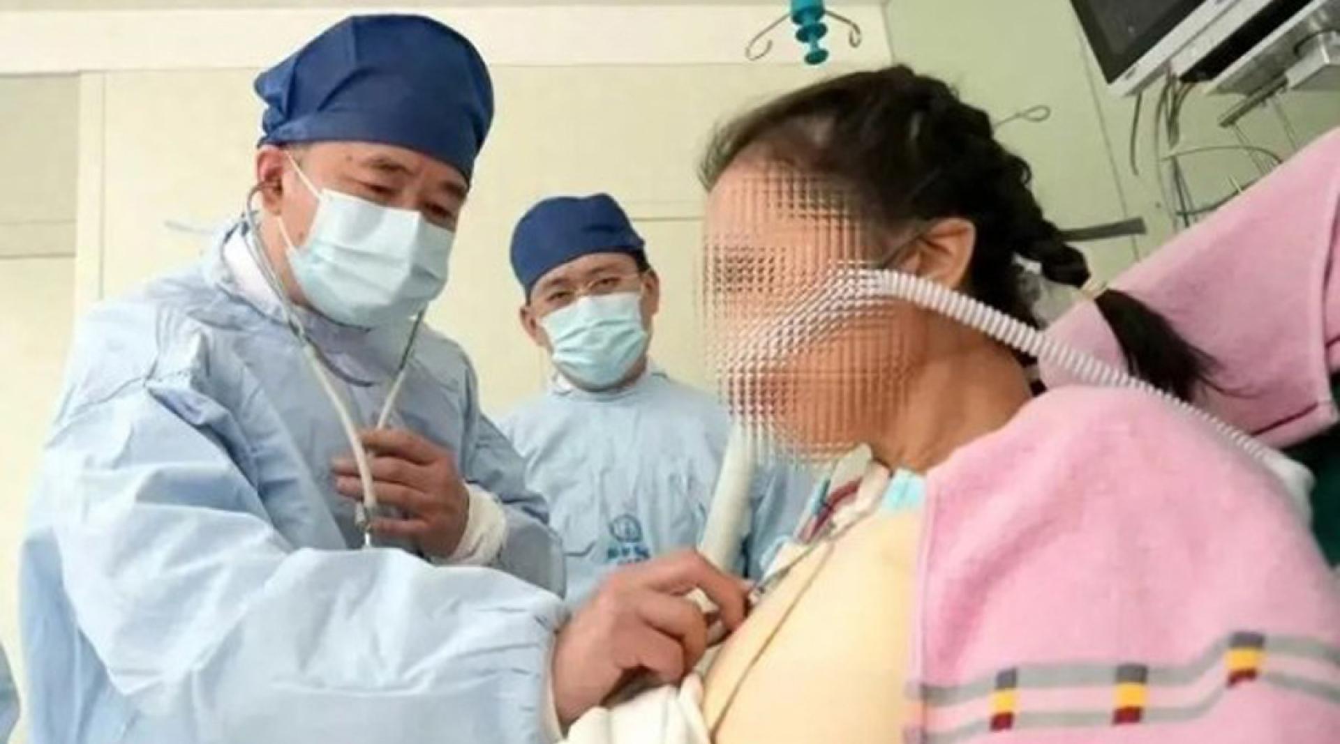
Four days after the surgery, Ms. Wu had been transferred to a general ward and could eat on her own and stand by the bedside for rehabilitation training. According to Dong Nianguo, conventional heart transplants require a 20 to 25 centimeter incision along the middle of the sternum. As Ms. Wu had been bedridden for a long time and was extremely weak, she could not withstand severe trauma. The new minimally invasive technique overturns traditional heart transplant approaches and surgical habits. While the difficulty of donor heart resuscitation is greatly increased, it does not damage the sternum or ribs, maximally preserving the integrity of the thoracic cage. This method is suitable for the vast majority of heart transplant patients, especially those with cachexia in end-stage heart failure, and their quality of recovery will be significantly better than conventional surgery patients.